
Figure 2. Comparison of glycinergic mIPSC properties in wild-type and spasmodic mice.
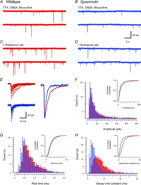
A and B, traces showing glycinergic mIPSCs recorded from wild-type and spasmodic SDH neurones in the presence of TTX (1 μm), CNQX (10 μm) and bicuculline (10 μm). Note the low mIPSC frequency in both wild-type and spasmodic recordings. C and D, bath application of ruthenium red (100 μm) increased mIPSC frequency in both wild-type and spasmodic neurones. E, overlain traces (captured from recordings in A and B) compare mIPSC properties in wild-type (upper left) and spasmodic (lower left) neurones (average appears as darker trace). A range of mIPSC amplitudes was observed in both genotypes, but mIPSC decay time was considerably faster in spasmodic mice. The overlain ‘averaged mIPSCs’, normalised to the same peak amplitude, further highlight the faster decay times in spasmodic mIPSCs. F–H, group data comparing mIPSC amplitude, rise time and decay time constant distributions. Cumulative probability plots are shown as insets. mIPSC amplitude was slightly reduced in spasmodic mice, rise times were similar for both genotypes, but decay time constants were significantly reduced in spasmodic neurones.