
Figure 1. Camera image of a typical cultured coronal slice used in these experiments.
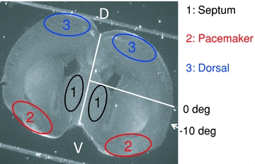
White lines indicate the midline of the slice and the 0 deg reference line extending from the midpoint of the midline to the pial surface. Numbered areas indicate the regions of interest (ROIs) used in Fluo-4 fluorescence records presented in the later figures (1: septal nuclei; 2: ventral cortical pacemaker; 3: dorsal cortex follower). The −10 deg label indicates the mean maximum extent of propagation of waves that do not enter the dorsal cortex. Waves that cross this point (fully propagating waves; FPWs) typically reach the dorsal midline, and never stop ventral to +60 deg (Conhaim et al. 2010). The same angular reference points were used on both sides of the slice.