
Figure 7. Electrical field potential recordings from the facial nucleus show respiratory activity in dorsal and lateral subnuclei.
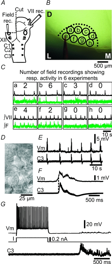
A, brainstem–spinal cord preparation, with one-side transverse cut through the mid-facial nucleus (oval), illustrating the position of nerve and field potential recording pipettes. VII, facial nerve; XII, hypoglossal rootlets; C1, first cervical nerve. B, photomicrograph of the live preparation, indicating the outline of the facial nucleus, eight recording positions (a–h), and the field potential recording pipette (labelled with red dye). D, dorsal; L, lateral; M, medial. C, integrated facial nerve, and field potential recordings (green traces) from positions a–h within the facial nucleus (∫VII, ∫F). Numbers in the middle of each box indicate the number of field potential recordings showing respiratory activity in 6 experiments. Note that the dorsal and lateral positions show respiratory field potential activity. D, contrast enhanced video micrograph of facial motoneuron. E, whole-cell patch recording of weak inspiratory drive potentials in a facial motoneuron (Vm), along with respiratory nerve activity on C3. F, cycle triggered average (6 cycles) of the membrane potential (Vm) and C3 activity from E. Dotted line indicates onset of nerve activity. G, firing pattern of a facial motoneuron in response to current (I) input, a single respiratory nerve burst on C3 and the resulting small inspiratory burst potential.