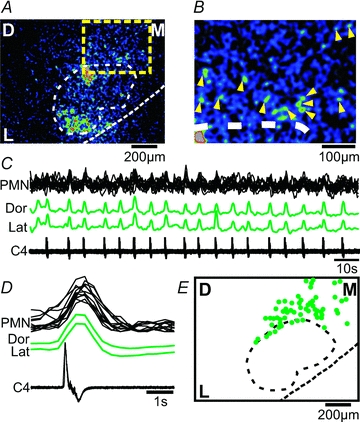
Figure 8. Putative facial premotoneurons, located dorsomedial to the facial nucleus, show respiratory activity.

A, CTA image from the facial nucleus triggered off respiratory activity on C4 (18 cycles) showing respiratory activity in the dorsal and lateral facial subnuclei, but also in cells located outside the facial nucleus (yellow square). B, zoom in of the yellow square region from A, showing individual cells (yellow triangles). C, respiratory activity on C4 and ΔF traces from the dorsal (Dor) and lateral (Lat) facial nucleus, and from individual premotoneurons (PMN, black overlaid traces). D, averages of respiratory activity (C4) and ΔF traces from 12 PMNs located dorsomedially to the facial nucleus. E, cumulated location of facial premotoneurons (green filled circles) showing respiratory activity (from 8 preparations). Note the predominantly dorsomedial positions of respiratory-modulated facial premotoneurons.
