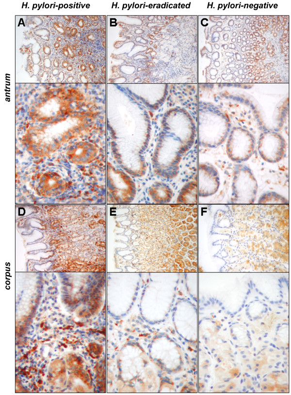
Figure 3.
Immunohistochemical detection of Progranulin in gastric antral mucosa. Immunohistochemical stainings exemplarily illustrate Progranulin expression in biopsies from gastric mucosa of the antrum and corpus, respectively, of H. pylori-positive (A+D), -eradicated (B+E) and -negative (C+F) subjects as identified in figure. Expression was seen in a granular pattern evenly distributed to the epithelial cytoplasm of the glands and crypts and accentuated to the base of the surface epithelium. Enlargements of panel A+D demonstrate the Progranulin-expressing immune cells (mostly granulocytes) in the mucosa of an infected individual, whereas these cells are less abundant in corresponding samples of H. pylori-eradicated (panel B+E) and -negative individuals (panel C+F). Microscope: Zeiss Axioscope 50, camera: Nikon coolpix 990; enlargements: ×100, ×400.

