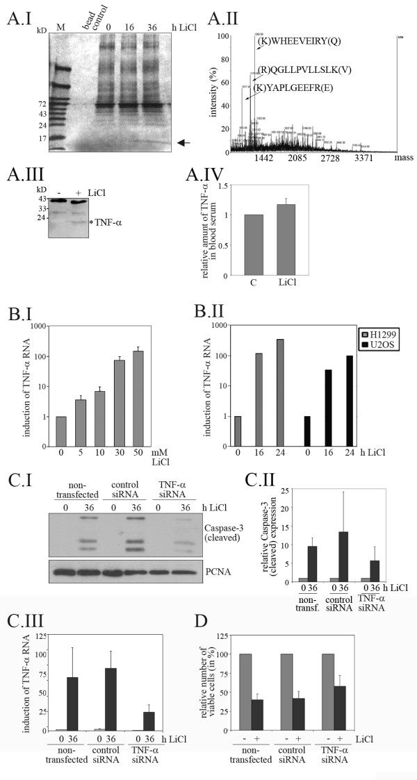
Figure 6.
LiCl stimulates the release of a TNF-α. (A.I) U2OS cells were cultured in 0.5% FCS and stimulated with 50 mM LiCl. At the indicated times, the culture medium was harvested and proteins were precipitated using Strataclean Resin. Proteins were eluted with sample buffer and separated on a 15% SDS-PAGE gel. The gel was silver-stained and photographed. The arrow points to a protein that is only present in LiCl treated cells. (A.II) The protein that was only present in the cell culture supernatant after treatment of cells with LiCl was eluted from the gel, digested with trypsin and subjected to MALDI-TOF sequencing. The graph shows the spectrogram from MALDI-TOF sequencing. Arrows point to peptides characteristic of TNF-α. (A.III) U2OS cells were cultured in 0.5% FCS and stimulated with 50 mM LiCl. After 36 hours, the culture medium was harvested and proteins were precipitated using Strataclean Resin, eluted with sample buffer and separated on a 15% SDS-PAGE gel. Proteins were transferred onto a PVDF membrane and probed with antibodies directed against TNF-α. (A.IV) Three Wistar rats were injected with 50 mg/kg of LiCl or with the vehicle (PBS) for 4 weeks. From 3 weeks after the first drug dose, blood was collected twice a week and TNF-α levels were determined in the serum using a rat-TNF-α ELISA kit (Invitrogen). Mean values and standard deviations were calculated from three bleedings and blotted. The value for rats that received the vehicle was set to 1. (B.I) U2OS cells were treated with the indicated concentrations of LiCl for 24 hours. RNA was prepared and the amount of TNF-α RNA was determined by qRT-PCR. Mean values and standard deviation of 2ΔCT values of 3 independent experiments were plotted. The data for untreated cells were set to 1. (B.II) U2OS and H1299 cells were treated with 50 mM LiCl and harvested at the indicated time points. RNA was prepared and the amount of TNF-α RNA was determined by qRT-PCR. Mean values of 2ΔCT values of 2 (H1299) and 3 (U2OS) independent experiments were plotted. The data for untreated cells were set to 1. (C) U2OS were transfected with siRNA targeted against TNF α or with a control siRNA, or left untransfected for control. 24 hours after transfection, 50 mM LiCl were added and the cells were incubated for a further 36 hours. Cells were harvested and divided into two aliquots. One of the aliquots was lysed and 50 μg of protein were separated on a 15% SDS-PAGE gel. Proteins were transferred onto a PVDF membrane and probed with antibodies directed against cleaved Caspase-3, and against PCNA for a control (C.I). Mean values and standard deviations from three independent experiments were calculated and plotted. Values for untreated cells were set to 1 (C.II). From the second aliquot, RNA was prepared and the amount of TNF-α RNA was determined by qRT-PCR. Mean values and standard deviations of 2ΔCT values of the three experiments were calculated and plotted. The values obtained for TNF-α RNA in the absence of LiCl were set to 1 (C.III). (D) U2OS cells were transfected with siRNA targeted against TNF-α or with a control siRNA, or left untransfected for control. 24 hours after transfection, 50 mM LiCl were added and the cells were incubated for a further 36 hours. Relative numbers of viable cells were determined by MTT-assay.

