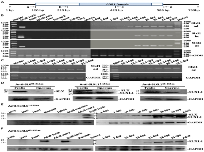
Figure 2. mRNA and protein expression of Slxl1 in mouse tissues.
(A). Positions of primers used in RT-PCRs. (B) Slxl1 mRNA was exclusively expressed in mouse testes indicated by RT-PCR analysis of multiple tissues; Slxl1 transcript was first detected at 14 days post partum (dpp) in postnatal mouse testes. (C) From 10.5 dpp to 22.5 dpp, Slxl1 transcript level increased gradually as detected by RT-PCR using the ad and ac primer sets. (D) One protein of about 25 kDa in adult testis was detected in Western blotting, however, the protein was not detected in the spermatozoa using anti-SLX95–212aa antibody. Using anti-SLXL11–155aa, two proteins of about 25 kDa and 18 kDa were detected in testes, while only the 18 kDa one was detected in spermatozoa. Only the 18 kDa protein was detected in either testis or spermatozoa using anti-SLXL165–155aa. (E–F) The expression of SLX and SLXL1 in mouse tissues and testes of different days post partum were detected using anti-SLXL11–155aa (E) and anti-SLXL165–155aa (F). mRNA and protein expression of GAPDH were used as internal controls for RT-PCR and Western Blot, respectively.

