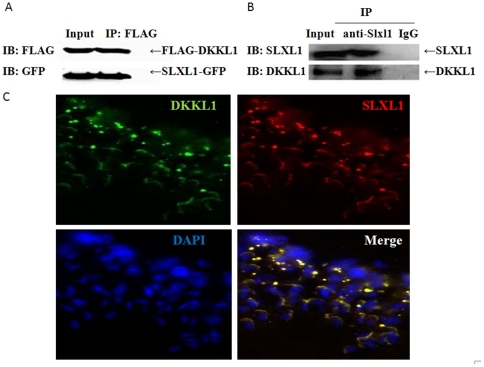
Figure 4. Co-immunoprecipitation and co-localization of SLXL1 and DKKL1.
(A). Coimmunoprecipitation of DKKL1-FLAG with SLXL1-GFP expressed in 293T cells. (B) Coimmunoprecipitation of DKKL1 with SLXL1 from testis lysates. Protein lysates were analyzed by standard Western blotting with anti-FLAG, anti-GFP, anti-SLXL1 and anti-DKKL1 antisera, respectively. (C) Co-localization of SLXL1 (red) and DKKL1 (green) in seminiferous tubules. The nuclei were stained with DAPI (blue).

