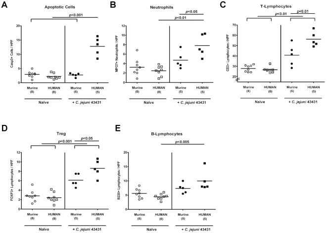
Figure 5. Immunopathological responses in the colon of “humanized” mice in situ following C. jejuni-infection.
Mfa and hfa mice were generated and orally infected with C. jejuni strain ATCC 43431 by gavage as described in methods section. The average numbers of apoptotic cells (postive for caspase-3, panel A), neutrophilic granulocytes (neutrophils, positive for MPO-7, panel B), T-lymphocytes (positive for CD3, panel C), Tregs (positive for FOXP3, panel D), and B-lymphocytes (positive for B220, panel E) from at least six high power fields (HPF, 400× magnification) per animal were determined microscopically in immunohistochemically stained colon sections of naïve (Naïve, open symbols) and infected (+C. jejuni ATCC 43431, filled symbols) mfa (circles) and hfa (squares) mice at day 12 p.i.. Numbers of analyzed animals are given in parentheses. Medians (black bars) and levels of significance (P-values) as compared to the indicated groups (determined by Student's t-test) are indicated. Data shown are representative for three independent experiments.

