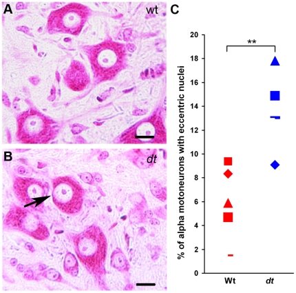
Figure 3. Increase in the number of eccentric alpha motor neuron nuclei in dt27J L1 spinal cord compared to wild type L1 spinal cord.
A. Normal alpha motor neurons were observed by light microscopy after Pyronine Y staining of the ventral horn of the L1 spinal cord from a wild type mouse. B. Eccentric motor nucleus (black arrow) observed in L1 spinal cord from a dt27J mouse. Scale bar, 10 µm (A, B). C. Dot plot graph showing the percentage of eccentric alpha motor neuron nuclei in the L1 spinal cord region as observed in individual wild type (n = 5) and dt27J (n = 5) mice (**p<0.01, student t-test).

