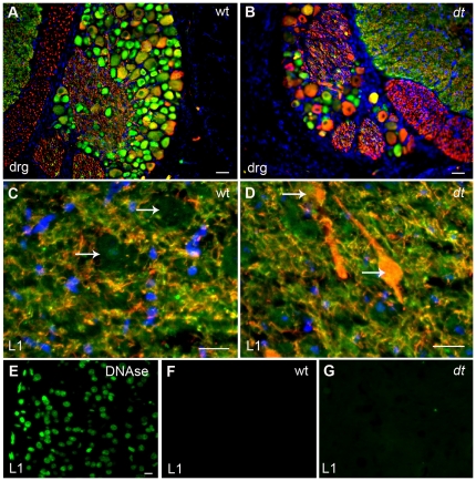
Figure 4. Analysis of neurofilament and alpha-internexin immunostaining in the dorsal root ganglion and ventral horn of the L1 spinal cord of wild type and dt27J mice.
A. Alpha-internexin staining (green) in cell bodies within the DRG of wild type mice. RT-97 staining of phosphorylated neurofilaments (red) is limited to a few cells. B. The DRG from dt27J mice is smaller and has fewer ganglion cells per DRG compared to wild type DRGs. RT-97 staining of phosphorylated neurofilaments (red) can be readily seen within the perikarya of many ganglion cells while alpha-internexin staining (green) is also present in a few cells. Scale bar, 10 µm (A, B). C. Ventral horn region of the L1 spinal cord from a wild type mouse showing no accumulation of phosphorylated neurofilaments within the perikarya of motor neurons (white arrows). Alpha-internexin staining (green) is observed throughout the dendrites and axons of the motor neurons. D. Ventral horn region of the L1 spinal cord from a dt27J mouse showing abnormal accumulation of phosphorylated neurofilaments within the perikarya (white arrows) of motor neurons. In comparison, alpha-internexin staining (green) is observed in dendrites and axons. Scale bar, 20 µm (C, D). E–G. TUNEL assay showed no labeling in cells in the L1 spinal cords of WT and dt27J mice (F–G). DNase treated L1 spinal cords were used as a positive control (E). Scale bar 10 µm.

