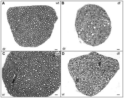
Figure 5. Defects in dt27J ventral motor and dorsal sensory spinal roots.
Toluidine blue staining of transverse sections of dorsal (dr) (A–B) and ventral (vr) roots (C–D). A. Dorsal sensory root from wild type mice showing many myelinated axons. B. Dorsal sensory root from dt27J mice showing several abnormalities including fewer axons, axons undergoing degeneration, and axonal swellings (sa). The dt dorsal sensory root is also smaller than the wild type counterpart. C. Ventral motor root from wild type mice showing many myelinated axons of different calibers. D. Ventral motor root from dt27J mice showing a mixture of myelinated and amyelinated axons of different calibers. Several large and intermediate caliber amyelinated axons are detected. The dt ventral motor root is smaller than the wild type counterpart and the axons are more compacted. Scale bar, 5 µm (all panels).

