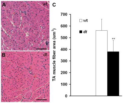
Figure 9. TA myofiber atrophy in dt27J mice at P15.
TA myofibers in cross-section were stained with hematoxylin and eosin. A. Healthy myofibers from TA wild type muscle were observed under light microscopy. B. Myofiber atrophy in TA dt27J muscle is observed. Myofibers are smaller and many more nuclei are visible. C. A graphical representation showing the TA muscle fiber area in µm2 from wild type (n = 6) and dt27J (n = 6) mice revealed muscle atrophy in dt27J mice (** p<0.01, student t-test). Scale bars (A, B), 100 µm.

