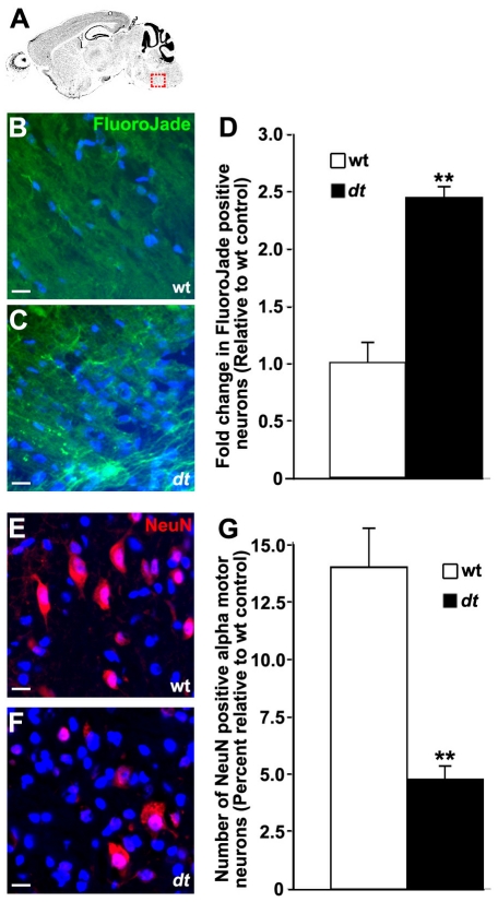
Figure 10. Motor neuron degeneration in the brainstem of dt27J mice.
Sagittal sections from mouse brainstem (trigeminal nerve of the pons) identified in (A) of P15 wild type (B) and dt27J (C) mice were stained for degenerating neurons with Fluorojade B. D. Fold change in neurodegeneration in dt27J brainstem relative to wild type littermates is depicted (** p<0.01, student t-test). Sagittal sections from mouse brainstem of P15 wild type (E) and dt27J (F) mice were antigenically labeled for mature neurons with NeuN. Alpha motor neurons are identifiable by their larger size (greater than 9 µm, arrow) relative to other neuronal cell types (arrowhead). Nuclei were counter stained with DAPI to facilitate quantification. G. Quantification of percent alpha motor neurons yielded a decrease in neuron number in the trigeminal nerve of dt27J mice relative to wild type littermates (**p<0.01, student t-test). Scale bars, 10 µm.

