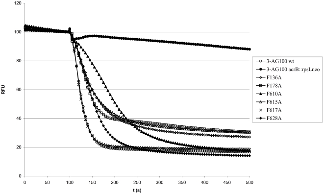
Figure 1. Representative 1,2′-DNA efflux curves of 3-AG100-derived AcrB binding pocket mutants.
After preloading with 4 µM 1,2′-DNA the cells were energized at 100 s with 50 mM glucose. Fluorescence intensity is given as relative fluorescence units (RFU) with preenergization levels adjusted to 100 RFU.

