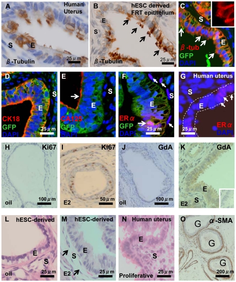
Figure 1. Characterisation of hESC-derived FRT epithelium in GF treated heterotypic (nMUM) recombinant 8 week xenograft.
β-tubulin expression in (A) ciliated simple columnar epithelia of human adult proliferative endometrium and (B) in hESC derived ciliated, simple columnar FRT epithelium (arrows indicates cilia). (C) immunofluorescence analysis of hESC derived FRT epithelium (green) where β-tubulin (yellow orange) is expressed on cell surface (inset shows a close up view of the cilia), the hESC origin of epithelial cells was assigned on the basis of GFP expression. Note that adjacent mouse uterine stromal cells are GFP−. hESC derived FRT epithelium co-localised (D) cytoplasmic CK18, (E) CA125 on epithelial surface (arrow), and (F) ERα (pink) with GFP+ hESC derived epithelial cells. Weak ERα expression was also evident in mouse uterine stromal cells (filled arrows). (G) ERα expression in human proliferative endometrial gland (full arrows), dotted line indicates epithelium. Ki67 expression in hESC derived FRT epithelium before (H) and after (I) estrogen injections showing estrogen-induced epithelial cell proliferation. The expression of GdA in hESC derived FRT epithelium without (J) and with (K) estrogen injections showing estrogen-induced cytoplasmic expression of GdA. H&E of hESC derived FRT epithelium lined by simple columnar cells without (L) and with (M) estrogen treatment, showing estrogen-induced increase in epithelial height and stromal oedema (arrows), classical hormonal responses of adult uterine epithelium (N). (O) Three hESC derived FRT epithelial structures surrounded by α-SMA positive cells. Inset in (K) is concentration matched mouse IgG1 negative control. Hoechst stained serial sections corresponding to (I) and (K) are found in Figure S6E–F. Abbreviations: α-SMA, α-smooth muscle actin; CA125, cancer antigen 125; E, epithelium; ERα, estrogen receptor alpha; E2, estrogen; G, gland; GdA, glycodelin A; S, stroma.

