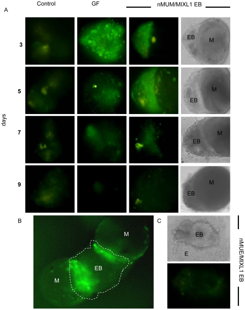
Figure 2. Neonatal mouse uterine mesenchyme induces MIXL1 expression in MIXL1GFP/w EBs.
(A) nMUM induced GFP expression in MIXL1 GFP/w EB recombinants after three days of co-culture (3rd panel), representative example of n>100. The duration of reporter activation is comparable to that observed in growth factor treated EBs (2nd panel, days 3,5), while little to no reporter activity was detected in the control (1st panel) (n>100) (×40 magnification, first 3 panels are fluorescent images, images in 4th panel represent phase contrast images of recombinants in 3rd panel). (B) Co-culture of two nMUM pieces (0.5 mm) with a larger EB (>4000 cells) activated reporter activity locally (dotted area is the EB) (×20 magnification). (C) No reporter activity was detected in embryoid bodies co-cultured with neonatal uterine epithelium (0.5 mm) (×40 magnification). Areas marked as mesenchyme in recombinants are based on morphological evidence and recombination experiments using ENVY hESC (refer to Figure S2A–C). Abbreviations: EB, embryoid body; E, neonatal mouse uterine epithelium; GF, growth factors; M, neonatal mouse uterine mesenchyme.

