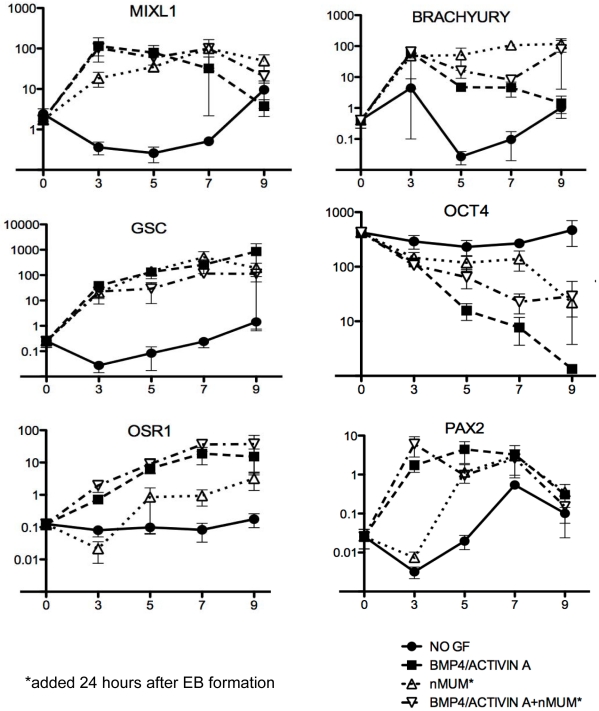
Figure 3. Neonatal mouse uterine mesenchyme induces expression of primary germ layer markers in ENVY/nMUM recombinants.
Real time PCR analysis of RNA collected from EB and recombinants cultured in serum-free BPEL medium. Growth factors (BMP4, 50 ng.ml−1 and ACTIVIN A, 20 ng.ml−1) were added immediately after forced aggregation of hESCs. Neonatal mouse uterine mesenchyme was added 24 hours after EB formation into either growth factor treated or untreated EB culture. Expression of genes relative to GAPDH were analysed by quantitative RT-PCR after 3, 5, 7 and 9 days incubation. Expression of target genes in undifferentiated hESCs is indicated as day 0 of differentiation (Data is plotted as mean±s.e.m., n = 3 independent differentiation experiments). Abbreviations: GF, growth factor; nMUM, neonatal mouse uterine mesenchyme.

