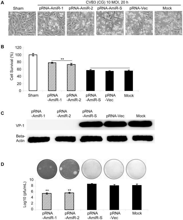
Figure 9. Anti-CVB3 (CG) effect of pRNA-AmiR chimerics in HL-1 cells.
HL-1 cells were transfected with different pRNA-AmiRs or mock-transfected with Oligofectamine™ only for 48 h and then infected with CVB3 (CG, 10 MOI 20 h) or sham-infected with PBS. Cell morphologies were analyzed by phase-contrast microscopy. Dying cells appeared rounding and detachment (A). Cell viability was measured by MTS assay (B), the viral VP-1 protein was detected by Western blot analysis (C) and viral particle formation was measured by plaque assay (D).

