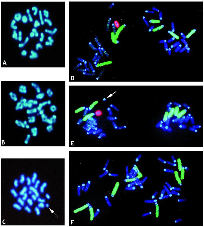
Figure 1.
Photographs of mouse MI and II spermatocytes after DAPI staining (A–C) and first-cleavage (1-Cl) zygotes after hybridization with chromosome-specific painting probes for chromosomes 1–3 and X (labeled with biotin and signaled with FITC) and chromosome Y [labeled with digoxigenin and signaled with rhodamine (D–F)]. Images were taken by using a Vysis (Downers Grove, IL) QUIPS Imaging Analysis System, and the final composite figure was made in Adobe PHOTOSHOP. (A) Normal MI spermatocyte. (B) MI spermatocyte with multiple chromosome structural aberrations. (C) MII spermatocyte with chromatid acentric fragments (one is indicated by the arrow, the second is in the center of the metaphase). (D) Normal 1-Cl zygote metaphase with the Y-bearing sperm-derived chromosomes on the left. (E) 1-Cl zygote with a centric fragment in the paternal chromosomes (arrow). (F) Hyperploid 1-Cl zygote. Note the presence ofa an extra chromosome (green) in the X-bearing sperm-derived chromosomes.

