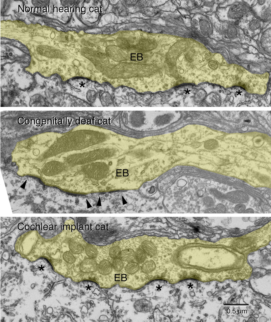
Figure 4.
Representative electron micrographs of endbulbs (EB, tinted yellow) taken from a normal hearing cat, a congenitally deaf cat, and a congenitally deaf cat stimulated electrically through a cochlear implant. The cats are matched in age. This series of micrographs illustrate that stimulation of the auditory nerve via a cochlear implant restores the structure of the synapse, specifically with respect to the size and curvature of the postsynaptic density (*) and the return of synaptic vesicle density adjacent to the synapse. (From Ryugo et al., 2005).

