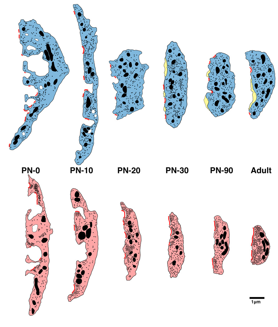
Figure 6.
Schematic of endbulb development in normal hearing (top) and deaf (bottom) cats. At birth, endbulb profiles have a highly convoluted membrane abutting the SBC, which gradually becomes smoother with age. Endbulbs of deaf animals are on average smaller than those found in age-matched normal hearing cats. The number of PSDs (red) at birth in deaf animals is less than half that of normal. Mature endbulbs of both deaf and normal animals have the same number of PSDs, the PSDs of deaf animals are longer and flatter than the normal convex PSDs. Deaf endbulbs exhibit an increase in synaptic vesicle (black dots) density near the PSDs. No differences were seen with respect to mitochondria (large black) size or volume fraction between the two groups. Endbulbs of normal cats begin to develop intermembraneous cisternae (yellow) around postnatal day 10, whereas endbulbs from deaf cats rarely develop them. (From Baker et al., 2010).

