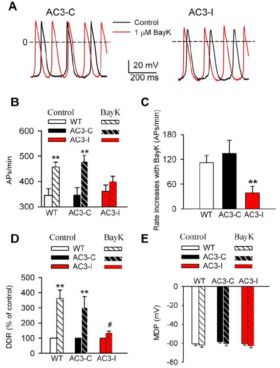
Fig 2.

SANC rate increases by BayK are reduced by CaMKII inhibition. A. Representative AP traces recorded before and after BayK in SANCs isolated from AC3-C and AC3-I mice. B. Spontaneous activity of SANCs in the absence and presence of 1 μM BayK. **P<0.01 vs control, n≥16 cells from more than 4 animals for each group. C. SANC rate increases by BayK in WT, AC3-C and AC3-I mice (data from panel B). ** P<0.001 vs WT or AC3-C, n≥16 cells from more than 4 animals for each group. D. BayK (1 μM) significantly increased the DDR in SANCs from WT and AC3-C, but not AC3-I mice. **P<0.01 vs control. # P<0.05 vs WT and AC3-C mice, n≥16 cells from more than 4 animals for each group. E. BayK did not significantly alter the MDP in any groups (data from panel D), n≥16 cells from more than 4 animals for each group.
