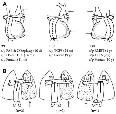
Fig. 5.
A: the anatomy of the cardiac mass, the conduit position and the azygous drainage. The main mass of ventricle is on the same side of the azygous drainage and the hepatic conduit position is inevitably on the contralateral side of the azygous drainage. B: the relationship between the conduit position and the azygous drainage site is related to the pattern of persistent pulmonary arteriovenous fistula after Fontan completion.28) PAB: pulmonary artery banding, COA: coactation of aorta, DS: Damus-Kaye-Stansel procedure, TCPS: total cavo-pulmonary shunt.

