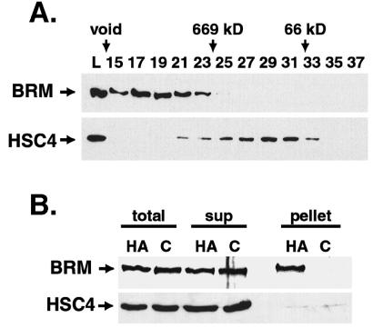
Figure 3.
HSC4 separation from the BRM complex in embryos. (A) Superose 6 gel filtration chromatography of embryo extracts, with fractions separated on Western blots and probed for BRM or HSC4. Lanes are designated by column fraction number or L, material loaded. Elution volumes of native molecular mass standards are indicated above. There is only a small amount of overlap between the BRM and HSC4 peaks. (B) BRM immunoprecipitates. An anti-HA mAb was used to immunoprecipitate extracts from wild-type control embryos (C) or embryos containing HA-epitope-tagged BRM protein (HA). Immunoprecipitation was from the pooled BRM peak fractions of a Superose 6 column. Western blotting of the starting material (total), supernatant (sup), and pellet samples revealed only trace amounts of HSC4 in both pellets, indicating no specific association with BRM.

