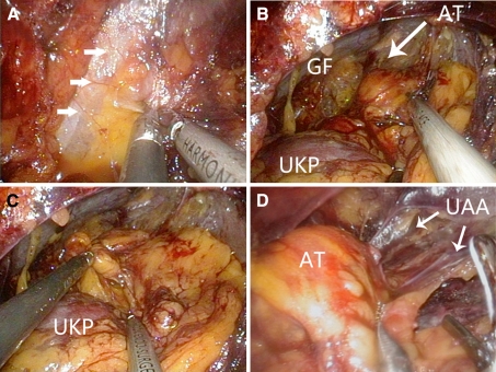
Fig. 2.
Internal views of the laparoendoscopic single-site retroperitoneoscopic adrenalectomy (LESS-ARA) procedure. A Gerota’s fascia is incised longitudinally along the posterior peritoneal reflection (white arrows) .B The adrenal tumor (AT) is identified in the first dissection plane between the perirenal fat and the anterior Gerota’s fascia (GF) located at the superomedial side of the upper kidney pole (UKP). C By grasping the periadrenal fat cephalad, the bottom of the adrenal gland or tumor is separated from the parenchymal surface of the upper kidney pole, after which the third dissection plane is developed. D The upper adrenal arteries (UAA) are transected

