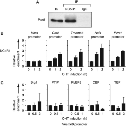
Figure 8.
Rapid recruitment of the NCoR1 complex to Pax5-binding sites of repressed target genes. (A) Interaction of Pax5 with the NCoR1 corepressor complex. Co-immunoprecipitation of Pax5 from nuclear extracts of Rag2–/– pro-B cells with NCoR1 antibodies. Pax5 was visualized in the immunoprecipitate (IP) by western blotting with a biotinylated rat anti-Pax5 mAb (detected with streptavidin-coupled horse radish peroxidase). Input (In; 1/200) and rabbit IgG were used as controls. (B) NCoR1 recruitment to repressed Pax5 target genes. KO-Pax5–ER pro-B cells were treated for the indicated time with 4-hydroxytamoxifen (OHT, 1 μM) before ChIP with an NCoR1 antibody. Input and precipitated DNA were quantified by real-time PCR with primer pairs amplifying the Pax5-binding regions of the indicated repressed Pax5 target genes and an inactive intergenic region on chromosome 1. The enrichment of precipitated DNA at the target sites relative to the chromosome 1 region was determined as described in the legend of Figure 6A. The relative enrichment at time point 0 was set to 1. The average values and standard deviations of two independent experiments are shown. The Hes1 promoter lacking Pax5-binding sites served as negative control. (C) No recruitment of activating protein complexes to the promoter of the repressed target gene Tmem66 upon Pax5–ER activation. KO-Pax5–ER pro-B cells were treated with OHT (1 μM) before ChIP with Brg1, PTIP, RbBP5, CBP and TBP antibodies and PCR analysis as described in (B). The average values and standard deviations of two independent experiments are shown.

