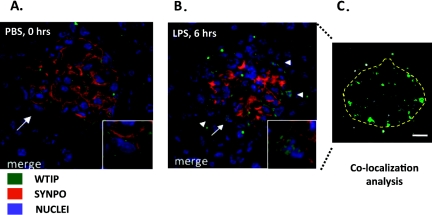
Fig. 4.
WTIP shifts into podocyte nuclei in a mouse model of nephrotic syndrome. (Panel A:) Glomerular co-staining of kidney cross sections of WTIP (green) and the podocyte-foot-process actin-binding protein synaptopodin (red). Nuclei are identified by TOPRO-3 (blue). Inset shows a zoom image of the podocyte marked by the arrow. WTIP and synaptopodin co-localize within podocyte foot processes, but no WTIP staining is seen within the podocyte nuclei. (Panel B:) Six hours after administration of LPS, WTIP has shifted into podocyte nuclei (inset), a finding confirmed with Image J analysis (shown in panel C). Image J identifies the spatial distribution of WTIP and TOPRO-3 pixels, and correlations above random overlap are pseudocolored green. The anatomic locations of the correlated pixels are consistent with podocyte nuclei. Used with permission (32).

