Fig. 1.
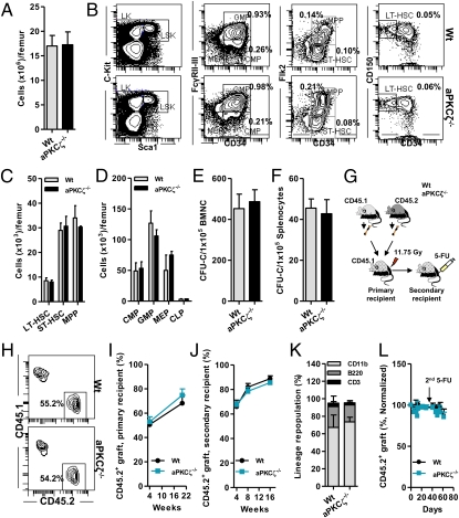
Deficiency of aPKCζ does not affect HSC activity or steady-state hematopoiesis. (A) Absolute numbers of nuclear cells present in the BM of WT or aPKCζ−/− mice (n = 3–4 mice per group). (B) Representative flow cytometry (FACS) contour diagram showing the frequency of hematopoietic stem cell and progenitors (HSC/P) present in the BM of WT or aPKCζ−/− mice. (C) Absolute numbers of HSCs present in the BM of WT or aPKCζ−/− mice (n = 3–4 mice per group). (D) Absolute numbers of hematopoietic progenitors (HPC) present in the BM of WT or aPKCζ−/− mice (n = 3–4 mice per group). (E) Absolute numbers of colony-forming progenitors (CFU-C) present in the BM of WT or aPKCζ−/− mice (n = 3–4 mice per group). (F) Absolute numbers of colony-forming progenitors (CFU-C) present in the spleen of WT or aPKCζ−/− mice (n = 3–4 mice per group). (A–F) Error bars represent SD. (G) Experimental set up: 2 × 10⁶ BM cells from CD45.2+ WT or aPKCζ−/− mice were mixed with 2 × 10⁶ CD45.1+ B6.SJLPtprca Pep3b/BoyJ WT BM cells and competitively (1:1 ratio) transplanted into lethally irradiated primary, secondary, and tertiary recipient mice and peripheral blood chimera was monitored at different time points. After 16 wk of engraftment, secondary recipient mice were further challenged (intraperitoneally) with two cycles (day 0 and day 40) of 5-FU injections (150 mg/kg body weight) and peripheral blood donor-derived chimera (CD45.2+) was monitored for 8 to 10 wk. (H) Representative FACS contour diagram showing competitive chimera of CD45.2+ WT or aPKCζ−/− cells serially transplanted into lethally irradiated CD45.1+ primary recipient mice from G. (I) Change in donor chimera (CD45.2+) in the peripheral blood of primary recipient mice (n = 6–8 mice per group). (J) Change in donor chimera (CD45.2+) in the peripheral blood of secondary recipient mice (n = 7 mice per group). (K) Frequency of lineage-repopulating myeloid (CD45.2+CD11b+) or B (CD45.2+B220+) or T (CD45.2+CD3+) cells present in the BM of the primary recipient mice (n = 6–8 mice per group). (L) Change in donor chimera (CD45.2+) in the peripheral blood of 5-FU–treated secondary recipient mice (n = 7 mice per group). (I–L) Error bars represent SEM.

