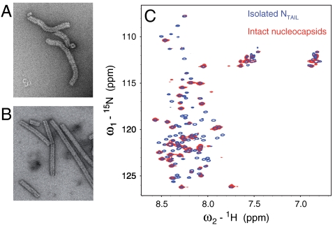
Fig. 3.
Electron microscopy and NMR studies of Measles virus nucleocapsids. (A) Electron micrograph (negative staining) of the 13C, 15N labeled nucleocapsid sample used for solution NMR studies. (B) Electron micrograph of trypsin-digested 13C, 15N labeled nucleocapsids. The solution NMR spectrum of this sample was empty. (C) Superposition of the 1H-15N HSQC spectrum of isolated NTAIL (blue) and intact nucleocapsids (red).

