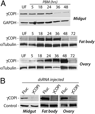
Fig. 1.
γCOPI protein expression in mosquito midgut, fat body, and ovary tissues. (A) Western blot analysis of γCOPI coatomer protein expression in midgut, fat body, and ovary tissue of blood fed WT female Ae. aegypti mosquitoes at various times postblood meal (PBM). Expressions of GAPDH and α-tubulin were used as protein-loading controls, with each lane containing protein extracts from an equivalent number of mosquitoes. (B) Knockdown of γCOPI coatomer protein expression in midgut, fat body, and ovary tissues of mosquitoes injected 3–4 d earlier with 400 ng γCOPI or Fluc dsRNA. Midgut tissues were collected before blood feeding, whereas fat body and ovary tissues were collected 24 h PBM. Western blotting was done using the antigen-specific γCOPI and either GAPDH antibody (midgut) or α-tubulin antibody (fat body and ovary) to control for protein loading.

