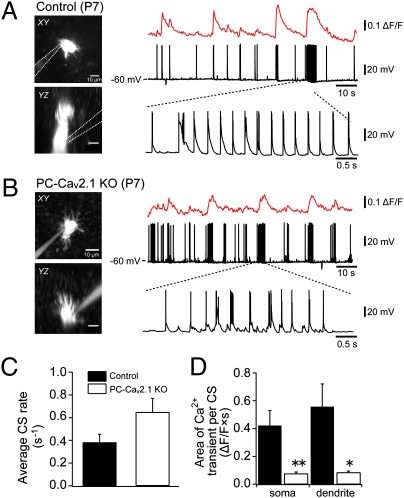
Fig. 2.
Reduction in CF-mediated Ca2+ transients in PCs of PC-Cav2.1 KO mice. (A and B) Simultaneous whole-cell recordings and two-photon Ca2+ imaging from PCs of control (A) and PC-Cav2.1 KO (B) mice in vivo. (Left) Horizontal (Upper) and parasaggital (Lower) projection images of recorded PCs at P7. (Right) Somatic Ca2+ transients (Top) and membrane potential (Middle) were recorded simultaneously. Note that trains of burst firing were often observed (Bottom). (C) Average firing rate of burst firing. (D) Somatic and dendritic Ca2+ elevation evoked by each train of burst firing was quantified as (area of Ca2+ transients)/(number of burst firings in train). *P < 0.002; **P < 0.0001.

