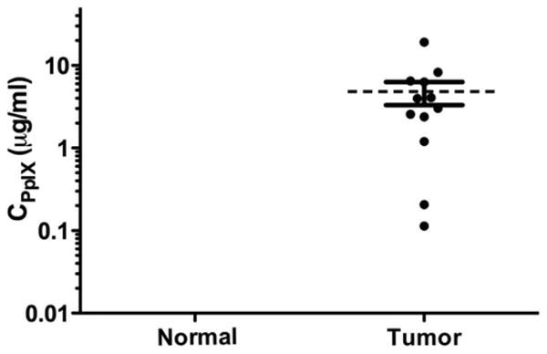
Fig. 2.

Protoporphyrin IX concentrations in skull base meningioma. Scatter plot of PpIX concentrations in normal and tumor tissue (mean 4.81 ± 1.49 μg/ml, range 0.11–19.15 μg/ml). Tumor tissue accumulated significantly more PpIX than normal tissue. Concentrations in normal dura were all below the level of quantification and outside the axis limits.
