Fig. 1.
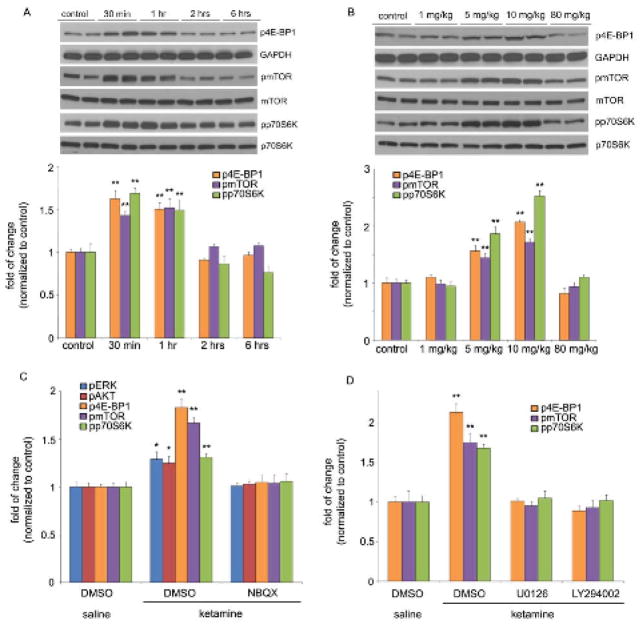
Ketamine transiently and dose-dependently activates mTOR signaling in rat prefrontal cortex (PFC). (A) Time course of ketamine (10 mg/kg, i.p.) induced mTOR signaling determined by Western blot analysis of phospho-mTOR (pmTOR), phospho-4E-BP1 (p4E-BP1), and phospho-p70S6K (pp70S6K) in synaptoneurosomes of PFC. Levels of total mTOR, GAPDH and p70S6K were also determined. (B) Dose-dependent activation, determined 1 hr after ketamine administration, of pmTOR, p4E-BP1 and pp70S6K. (C) Pre-treatment (10 min) with NBQX (10 mg/kg, i.p.) blocked ketamine (10 mg/kg, i.p.) activation of pmTOR, p4E-BP1, and pp70S6K, as well as upstream signaling kinases phospho-ERK (pERK) and phospho-Akt (pAkt) (analyzed 1 hr after ketamine). Levels of pERK1 and pERK2 were similarly regulated and were combined for quantitative analysis. (D) Pre-treatment (30 min) with inhibitors of ERK (U0126, 20 nmol, ICV) or PI-3k/Akt (LY294002, 20 nmol, ICV) abolished ketamine (10 mg/kg, i.p.) activation of mTOR signaling proteins (analyzed 1 hr after ketamine administration). Values represent mean ± SEM [n = 4 animals; * P < 0.05; ** P < 0.01, Analysis of Variance (ANOVA)].

