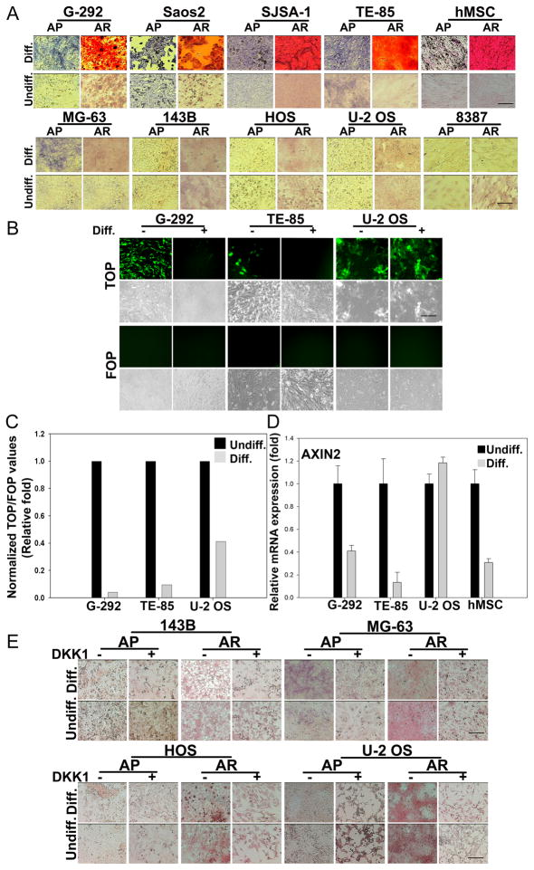
Figure 7. In vitro differentiation properties of Wnt activated human sarcoma cells.
(A) Sarcoma lines were plated at 10,000 cells/well in 12-well plates and exposed to osteogenic differentiation media (Diff.) for two weeks followed by fixation and staining for either alkaline phosphatase (AP, blue) or Alizarin Red S (AR, red). Control cells were maintained in basal medium (undiff.).
(B) Sarcoma lines stably expressing TOP- or FOP-GFP reporters were cultured as described in (A) and images were captured with a fluorescence microscope or by phase contrast.
(C) TOP-luciferase reporter activity in sarcoma cells cultured in basal or osteogenic differentiation media. Values are normalized to Renilla. Normalized values obtained for cells cultured in basal media (undiff.) were set at 1.
(D) Real-time PCR measurement of AXIN2 using total RNA extracted from sarcoma cells or hMSCs cultured in the presence or absence of osteogenic medium for two weeks as described in Fig. 2C. Normalized values obtained for cells cultured in basal media (undiff.) were set at 1. Error bars indicate SD of mean values from triplicates and are representative of two independent experiments.
(E) Sarcoma lines resistant to osteogenic differentiation were cultured in the presence or absence of osteogenic medium along with DKK1 (100ng/ml) added every other day for two weeks and stained for AP and AR.
Scale bars indicate 100μm. See also Figure S6.

