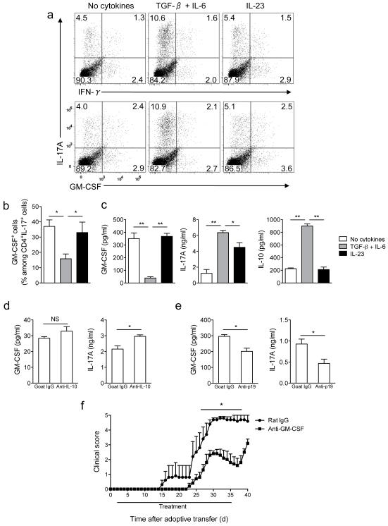
Figure 4. Neutralization of GM-CSF attenuates adoptive EAE.
(a) Splenocytes from MOG35-55-immuzed C57BL/6 mice were activated with MOG35-55 (20 μg/ml) in the presence of TGF-β plus IL-6, IL-23 or without added cytokines. Cells were then stimulated with PMA and ionomycin in the presence of GolgiPlug, stained and analyzed by flow cytometry. CD4+ cells are shown. (b) Percentage of GM-CSF+ cells among CD4+IL-17A+ cells after the second stimulation. (c) GM-CSF, IL-10, and IL-17A concentrations in cell culture supernatants measured by ELISA. (d) GM-CSF and IL-17A concentrations in supernatants of cell cultures stimulated as in (a) in the presence of TGF-β plus IL-6 and treated with anti-IL-10 or goat IgG. (e) GM-CSF and IL-17A concentrations in supernatants of cell cultures stimulated as in (a) without added cytokines and treated with anti-IL-23p19 or goat IgG (f) Clinical scores of irradiated wild-type recipient mice that received 5×10⁶ CD4+ cells enriched from EAE splenocytes activated in the presence of IL-23. Mice were treated with either anti-GM-CSF or rat IgG from day 2 to day 35 post cell transfer. *p< 0.01; **p < 0.001. Data are representative of two (d, e and f) or four (a, b, and c) independent experiments (error bars, s.e.m).

