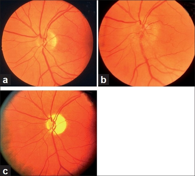
Figure 5.

Fundus photographs of left eye of a 53-year-old man: (a) Normal disc before developing NA-AION, (b) with optic disc edema and hyperemia during the active phase of NA-AION, and (c) after resolution of optic disc edema and development of optic disc pallor (more marked in temporal part than nasal part)[16]
