
Figure 6.
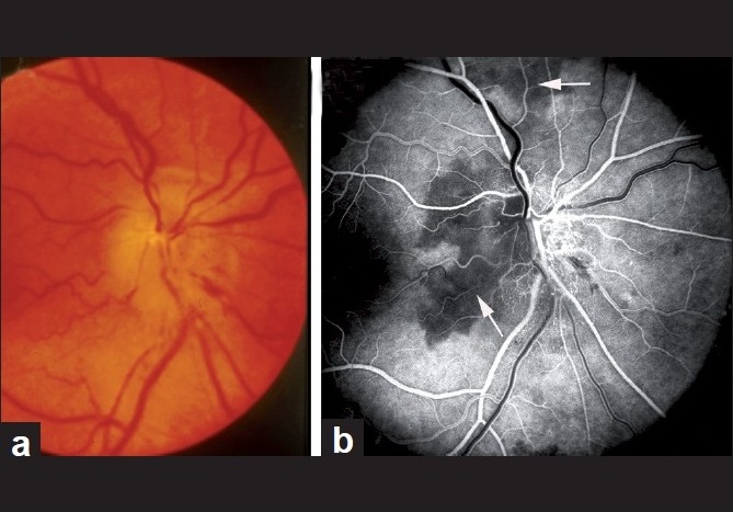
Fundus photograph (a) and fluorescein fundus angiogram (b) of right eye with NA-AION. (a) Optic disc edema, hyperemia and hemorrhages on optic disc; (b) fluorescein fundus angiogram shows non-filling of temporal part of the peripapillary choroid (arrow) and adjacent optic disc and the choroidal watershed zone (arrow)[36]