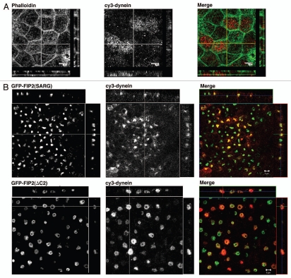
Figure 1.
Rab11-FIP2 mutants alter the localization of the microtubule motor protein, dynein heavy chain. Confocal fluorescence microscopic images of polarized MDCK cells. X-Y plane images are shown flanked by X-Z projections (horizontal) and Y-Z projections (vertical). (A) Parental T23 MDCK cells were stained with Alexa-488-phalloidin (pseudo-colored green) and antibodies against dynein (pseudo-colored red) and imaged by confocal microscopy. Dynein was observed in association in vesicles at the periphery of the cells, especially at the lateral borders. (B) T23 MDCK cells stably expressing either EGFP-Rab11-FIP2(SARG) or EGFP-Rab11-FIP2(ΔC2) were stained with antibodies against dynein and imaged by confocal microscopy. Expression of both Rab11-FIP2 mutants caused accumulation of dynein within the collapsed membrane cisternae. Images were taken with a 100x lens. Scale bars represent 5 µm.

