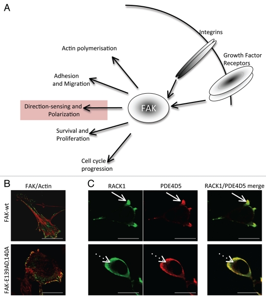
Figure 1.
(A) Schematic showing the recognized cellular functions of FAK, highlighting the direction-sensing and polarization functions we describe here. (B) Images show comparison of cell shape and actin filaments in FAK-deficient SCC cells re-expressing either FAK-wt or the FAK/RACK1 binding impaired mutant FAK-E139A,D140A. (C) The co-staining of RACK1 and PDE4D5 is shown in protrusive nascent adhesions as FAK-wt SCC cells are plated on to FN for 15 min (solid arrows). By comparison, both RACK1 and PDE4D5 are cytoplasmic in FAK-E139A,D140A-expressing cells (broken arrows). Scale bars 20 µm.

