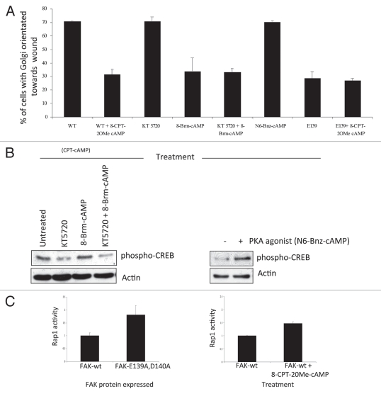
Figure 2.
(A) FAK-wt or FAK-E139A,D140A cells (as indicated) were plated on FN for 2 hours and wounded in the presence of 8-CPT-2OMe cAMP (CPT-cAMP; 10 µM), KT 5720 (1 µM), 8-Brm-cAMP (10 µM), KT 5720 + 8-Brm-cAMP or N6-Bnz-cAMP (10 µM). After 1.5 hours cells were fixed and stained with the Golgi marker anti-GM130, TRITC phalloidin and DAPI, to assess polarization in response to the wounded areas. The percentage of each cell type with the Golgi orientated to the wound was calculated by counting 100 cells in 3 experiments and is shown in the graph. (B) FAK-wt cells were treated with KT 5720 (1 µM) and/or N8 = Brm-cAMP (left parts) and with 6-Bnz-cAMP (10 µM) for 30 min then immunoblotted using anti-phospho-CREB and actin antibodies as probes. (C) FAK-wt cells with or without CPT-cAMP (10 µM for 3.5 hrs, time point chosen to maintain consistency with cell polarization experiments, conditions detailed above; left part) or FAK-wt and FAK-E139A,D140A cells (right part), were harvested and a Rap1 assay performed as described in Materials and Methods. Quantification of Rap1-GTP normalized to total Rap1 from three separate experiments is shown.

