
Fig. 1.
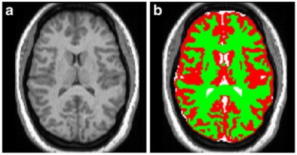
MPRAGE image segmentation. Individual MPRAGE images are registered to standard space before segmentation is accomplished. a Normalised MPRAGE image. b Segmented image: grey matter (red), white matter (green) and CSF (white) partitions are overlaid on top of the normalised MPRAGE image