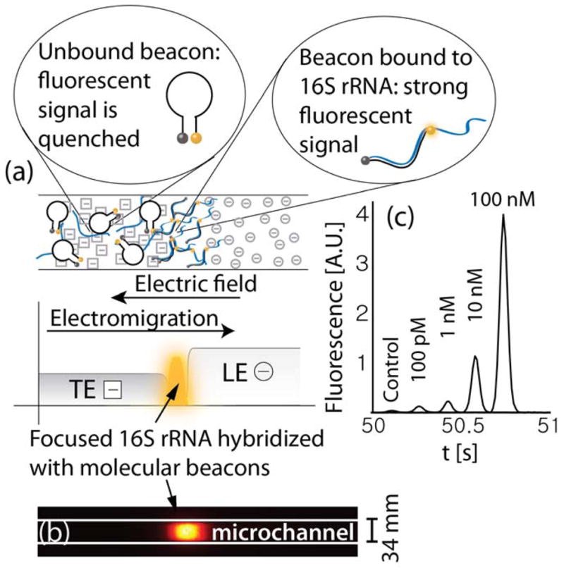
Figure 1.

(a) Schematic showing simultaneous isotachophoretic extraction, focusing, hybridization (with molecular beacons), and detection of 16S ribosomal RNA bound to a molecular beacon. Hybridization of the molecular beacon to 16S rRNA causes a spatial separation of its fluorophore and quencher pair resulting in a strong and sequence-specific increase in fluorescent signal. (b) Raw experimental image showing fluorescence intensity of molecular beacons hybridized to synthetic oligonucleotides using ITP. (c) Detection of oligonucleotides having the same sequence as the target segment of 16S rRNA. Each curve presents the fluorescence intensity in time, as recorded by a point detector at a fixed location in the channel (curves are shifted in time for convenient visualization). 100 pM of molecular beacons and varying concentrations of targets were mixed in the trailing electrolyte reservoir. The total migration (and hybridization) time from the on-chip reservoir to the detector was less than a minute.
