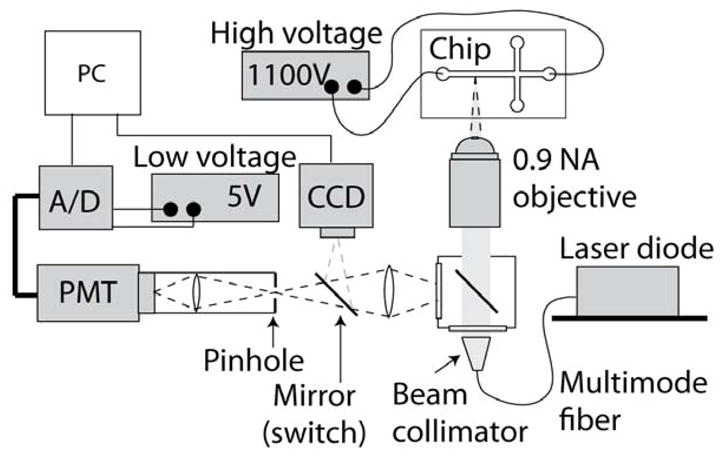
Figure 2.

Schematic of the experimental set-up with pointwise confocal optics. We used a 0.9 numerical aperture water-immersion objective to collect the light emitted by the molecular beacons within the microfluidic chip. A 400 μm pinhole was placed at the image plane, allowing collection of light from within the 12 μm deep channel, while rejecting out-of-focus light. The light was refocused onto a PMT for detection. Excitation is performed using a variable-power laser diode coupled into the illumination port of the microscope using a multimode optical fiber. The beam is expanded and collimated before being focused onto the channel using the same objective used for light collection. A CCD camera is used for alignment of the laser and microchannel prior to each experiment.
