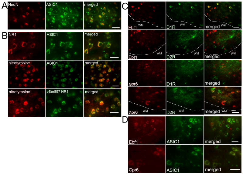
Figure 2.
Double immunofluorescence of ASIC1 and NeuN in normal pig striatum (A); ASIC1 and NMDA receptor NR1 subunit or nitrotyrosine, and nitrotyrosine and phosphorylated NR1 Ser897 (pSer897 NR1) in pig putamen at 3 h of recovery after H-I (B); Ebf1 or Gpr6 and dopamine D1 receptor (D1R) or dopamine D2 receptor (D2R) in pig putamen at 3 h of recovery after H-I (C); ASIC1 and Ebf1 or Gpr6 in normal pig striatum (D). White dash line indicates the boundary between grey matter and white matter. White matter (WM). Scale bar = 20 μm.

