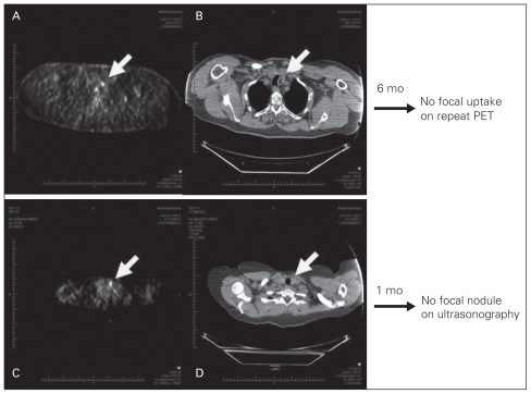
Fig. 3.
(A) Follow-up fluorodeoxyglucose positron emission tomography (FDG-PET) scan demonstrated 1.0 cm, 4.2 maximum standardized uptake value (SUV), uptake in the left lobe of the thyroid in a 75-year-old man with lymphoma. (B) Computed tomography also showed incidental thyroid nodule corresponding to FDG-PET. Repeat FDG-PET performed 6 months later, however, failed to show any uptake in the thyroid. (C) Follow-up FDG-PET scan demonstrated 1.0 cm, 4.5 maximum SUV, uptake in the left lobe of the thyroid in a 50-year-old woman with sarcoma. (D) Computed tomography also showed incidental thyroid nodule corresponding to FDG-PET. Subsequent ultrasonography 1 month later, however, did not show any focal lesion in the thyroid.

