
Figure 1.
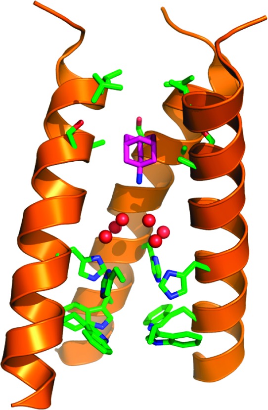
Structure of amantadine (purple carbon atoms) in complex with the transmembrane domain of M2, as determined by solid-state NMR9 (PDB: 2KQT), with the water structure seen in the high-resolution crystallographic structure (PDB: 3LBW) superimposed. Six water molecules from 3LBW, shown as red spheres, lie above the four His37 and Trp41 residues shown near the bottom of the structure (side chains shown in stick). The apolar region of the drug projects into the cavity formed by Val27, Ser31, and Ala30 (near the upper portion of the structure), while the ammonium group projects downward toward the water cluster. One helix has been removed for clarity.