
Figure 5.
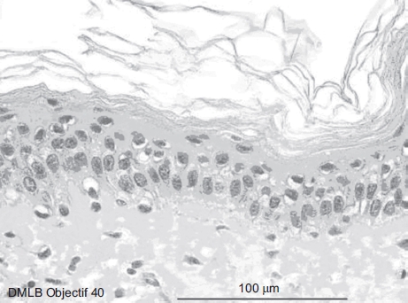
Skin histological aspect at 2 min after a 20-sec exposure to 30 μL of 70% hydrofluoric acid (HF). The skin showed four to five cellular layers with definitely abnormal morphology: cells with nuclei becoming pyknotic, especially in the higher epidermal layers, and the cytoplasm becoming acidophilic as reflected by orange keratinocyte pigmentation. The cellular structures in the dermis showed normal morphology. (See colour version of this figure online at www.informahealthcare.com/cot)