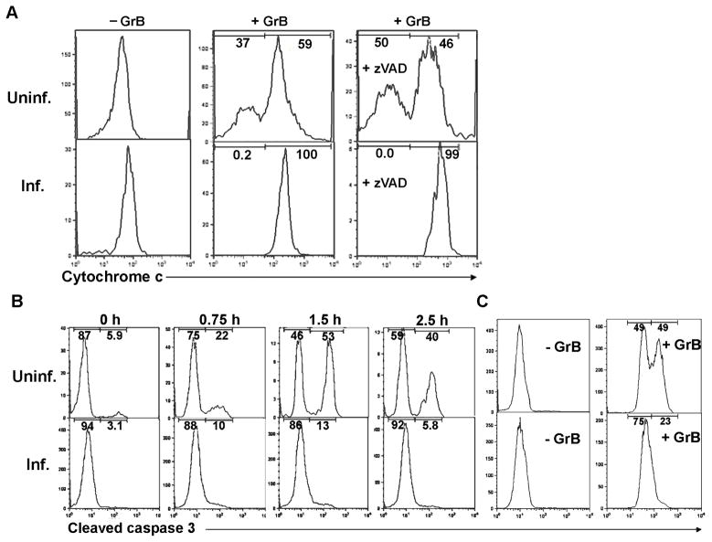
Fig. 3.
Inhibition of granzyme B (GrB)-induced cytochrome c release and caspase activation in Toxoplasma gondii-infected cells. (A) Jurkat cells were infected with T. gondii expressing yellow fluorescent protein (YFP-RH) at a multiplicity of infection (MOI) of 1 for 16 h and treated for 2.5 h with 6 ng/ml listeriolysin O (LLO) in the presence or absence of 60 nM GrB. Some samples received 100 μM n-benzyloxycarbonyl-val-ala-asp-fluoromethyl ketone (zVAD) 1 h before LLO addition. Cytochrome c release was measured by flow cytometry following digitonin permeabilization and cytochrome c immunostaining to determine remaining cell-bound cytochrome c. Infected (Inf.) and uninfected (Uninf.) fractions of the same culture were analyzed. The frequency of infection was 16.3%. (B) Jurkat cells were infected with YFP-RH at a MOI of 1 for 16 h and treated for the indicated times with 6 ng/ml LLO and 60 nM GrB. Fixed cells were immunostained to detect cleaved caspase-3 and analyzed by flow cytometry. Infected and uninfected fractions of the same culture were analyzed. The frequency of infection was 81%. (C) HeLa cells were infected with YFP-RH at a MOI of 4 for 16 h, treated with 2.25 μg/ml streptolysin O and 60 nM GrB for 1.5 h and then analyzed as in (A). The frequency of infection was 41.5%. The data in the figure are representative of two to three experiments.

