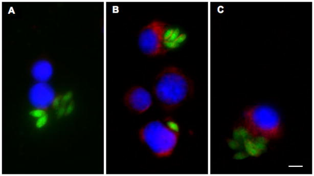
Fig. 7.
Uptake of granzyme B (GrB) in Toxoplasma gondii-infected cells. Jurkat cells were infected with T. gondii expressing yellow fluorescent protein for 16 h and treated for 1 h with 6 ng/ml listeriolysin O in the absence (A) or presence (B,C) of 60 nM GrB. Cells were fixed either immediately (A,B) or after trypsin treatment (C) and stained to detect GrB (red), followed by counterstaining with DAPI (blue). Infection is indicated by the presence of parasitophorous vacuoles detected as clusters of green parasites. Scale bar = 5 μm. The results are representative of three similar experiments.

